eo_logo
 
Product added to cart

4X Objective, Nikon CFI Plan Fluor

See More by Nikon

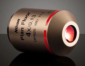
4X Objective, Nikon CFI Plan Fluor, #88-378

×
Stock #88-378 Contact Us
×
Quantity Selector - Use the plus and minus buttons to adjust the quantity. +
₹39,585
Qty 1+
₹39,585
Volume Pricing
Request Quote

Specifications

General

Model Number:
MRH00045
Compatible Tube Lens Focal Length (mm):
Focal Length: 200mm
Type:
Microscope Objective
Style:
Infinity Corrected
Manufacturer:
Nikon

Physical & Mechanical Properties

Field of View (mm):
6.25
Length excluding Threads (mm):
42.70
Maximum Diameter (mm):
30
Weight (g):
140.00

Optical Properties

Compatible Cover Glass Thickness (mm):
N/A
Horizontal Field of View, 1/2" Sensor:
1.60mm
Horizontal Field of View, 2/3" Sensor:
2.20mm
Magnification:
4X
Numerical Aperture NA:
0.13
Working Distance (mm):
17.2
Field Number (mm):
25
Parfocal Length (mm):
59.9
Immersion Liquid:
N/A

Sensor

Maximum Sensor Format:
2/3"

Threading & Mounting

Mounting Threads:
M25 x 0.75

Regulatory Compliance

RoHS 2015:
Certificate of Conformance:
Reach 235:

Product Details

  • Provide High Contrast Fluorescence Observation
  • Ideal for Multiple Microscopy Techniques
  • High Transmission from UV to the IR

Nikon CFI Plan Fluor Objectives feature high transmission from the ultraviolet to the infrared, along with exceptional flatness across the entire field of view. These objectives are ideal multipurpose objectives for a variety of microscopy techniques, including brightfield, fluorescence, polarizing, or DIC microscopy. These microscope objectives are suitable for high contrast fluorescence observation or for applications including photomicrography. Nikon CFI Plan Fluor Objectives have M25 x 0.75 mounting threads. The objectives are available in magnifications from 4X up to 60X.  
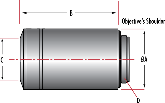
Technical Information

Dimensions (mm)
Stock No. Nikon Model Number Immersion Liquid Magnification Parafocal Length
A B C D
#88-378 MRH00041

N/A

4X 60.06 30.0 42.7 16.5 M25 x 0.75
#88-379 MRH00101 N/A 10X 60.06 30.0 42.4 18.0 M25 x 0.75
#38-004 MRP07220 Water 16X 75.0 35.0 72.0 6.0 M32 x 0.75
#88-380 MRH00201 N/A 20X 60.06 28.0 57.8 8.5 M25 x 0.75
#88-381 MRH00401 N/A 40X 60.06 30.0 59.2 10.0 M25 x 0.75
#88-382 MRH00602 N/A 60X 60.06 31.5 59.2 5.0 M25 x 0.75

Related Products

Frequently Purchased Together

Resources

Filter

Understanding Microscopes and Objectives

Learn about the different components used to build a microscope, key concepts, and specifications at Edmund Optics.

View Now

Multiphoton Microscopy

Multiphoton microscopy is ideal for capturing high-resolution 3D images with reduced photobleaching and phototoxicity compared to confocal microscopy.

View Now

Confocal Microscopy

Confocal microscopy provides high resolution, elimination of out-of-focus glare due to spatial filtering, and reduction of light-induced damage to the sample.

View Now

Light Sheet Microscopy

Light sheet fluorescence microscopy uses a 2D laser sheet to illuminate a thin slice of the sample and excite fluorescence, reducing phototoxicity and damage.

View Now

Optical Microscopy Application: Fluorescence

Wondering how fluorescence microscopy works? Find out about the technique, systems, and more at Edmund Optics.

View Now

Fluorescence Microscopy

Infinite Conjugate Tube Length

Fluorophores and Optical Filters for Fluorescence Microscopy

Want to know more about fluorophores and optical filters for fluorescence microscopy? Find out more information and in stock optical filters at Edmund Optics.

View Now

Understanding Infinity Corrected Objective Resolving Power and Magnification

Microscopes are used in a variety of fields and applications. To understand how resolving power, magnification, and other aspects work, read more at Edmund Optics.

View Now

Using Tube Lenses with Infinity Corrected Objectives

Want to use an infinity corrected objective to make an image? You will need a tube lens to do it! Find out why and how it works at Edmund Optics.

View Now

What is an "in-line" microscope?

What is an infinity-corrected objective?

Can I use an infinity-corrected objective in my microscope to get a longer working distance?

What is "DIN" and "JIS"? What is the difference between them?

I’m using the MT-1 Accessory Tube Lens and MT-1/MT-2 C-Mount Adapter. Two thin brass rings came with the adapter. How are they used?

What is the significance of the field number of an objective and what is it dependent on?

Plan Objective

Semi-Plan Objective

Tube Lens

Fluorescence Imaging with Laser Illumination

Fluorescence imaging systems are composed of three major components, an illumination source, a photo-activated fluorophore sample, and detector.

View Now

Fluorescent Filter

The Future of Microscope Objectives

Advances in Microscopy Tackle the Challenges of the Future

View Now

Optical Microscopy Application: Brightfield Illumination

Looking to use a simple technique for optical microscopy? Check out this article on brightfield illumination to find out more at Edmund Optics.

View Now

Optical Microscopy Application: Darkfield Illumination

Darkfield illumination is the opposite of brightfield illumination. Find out how darkfield differs from brightfield in optical microscopy at Edmund Optics.

View Now

Optical Microscopy Application: Phase Contrast

Think you know all the advantages for using phase contrast in optical microscopy? Advantages, image appearance, and technical details can be found at Edmund Optics.

View Now

What is the difference between Achromatic, Semi-Plan and Plan objectives?

You have a variety of Mitutoyo Tube Lenses (MT-4, MT-1, and MT-2). How can you connect them to a Mitutoyo objective and C-mount camera?

I have an MT-L Accessory Tube Lens but I am unsure about mounting it to my objective and C-Mount camera. Can you explain how it works?

Achromatic Objective

Apochromatic

Confocal Microscopy

DIN (Deutsche Industrie Norm)

Periplan Eyepiece

Optical Cage System Application: Digital Video Microscope

Are you looking to simplify the assembly of a digital video microscope? Find out how a TECHSPEC Optical Cage System allows for quick build at Edmund Optics.

View Now

Optical Microscopy Application: Differential Interference Contrast

Differential interference contrast (DIC) is one of the polarization techniques that can be used in optical microscopy. Learn about this technique at Edmund Optics.

View Now

Field Number

Optical Cage System Application: Differential Interference Contrast (DIC) Digital Microscope

Differential interference microscopy enhances the contrast of object features that are otherwise difficult to observe using brightfield microscopy.

View Now

Darkfield Illumination

Diffraction Limit

Parfocality

Digital Video Microscope Objective Setups

Digital video microscopes use a camera to capture and record images. Read more about the components needed to assemble a video microscope at Edmund Optics.

View Now

Brightfield Illumination

Field of View (FOV)

Magnification

Numerical Aperture (NA)

Fluorescence Microscopy: In-Line Illumination with Imaging Filters

Want to know about fluorescence microscopy? Read this article by a Biomedical Product Line Engineer at Edmund Optics to learn more.

View Now

Refraction

 
Sales & Expert Advice
1-800-363-1992
or view regional numbers
Easy-to-Use
QUOTE TOOL
enter stock numbers to begin