Engineers are available to assist.


In Vivo Fluorescence Imaging from UVP has Important Implications for Cancer Research, Heart Disease, and Gene Therapy

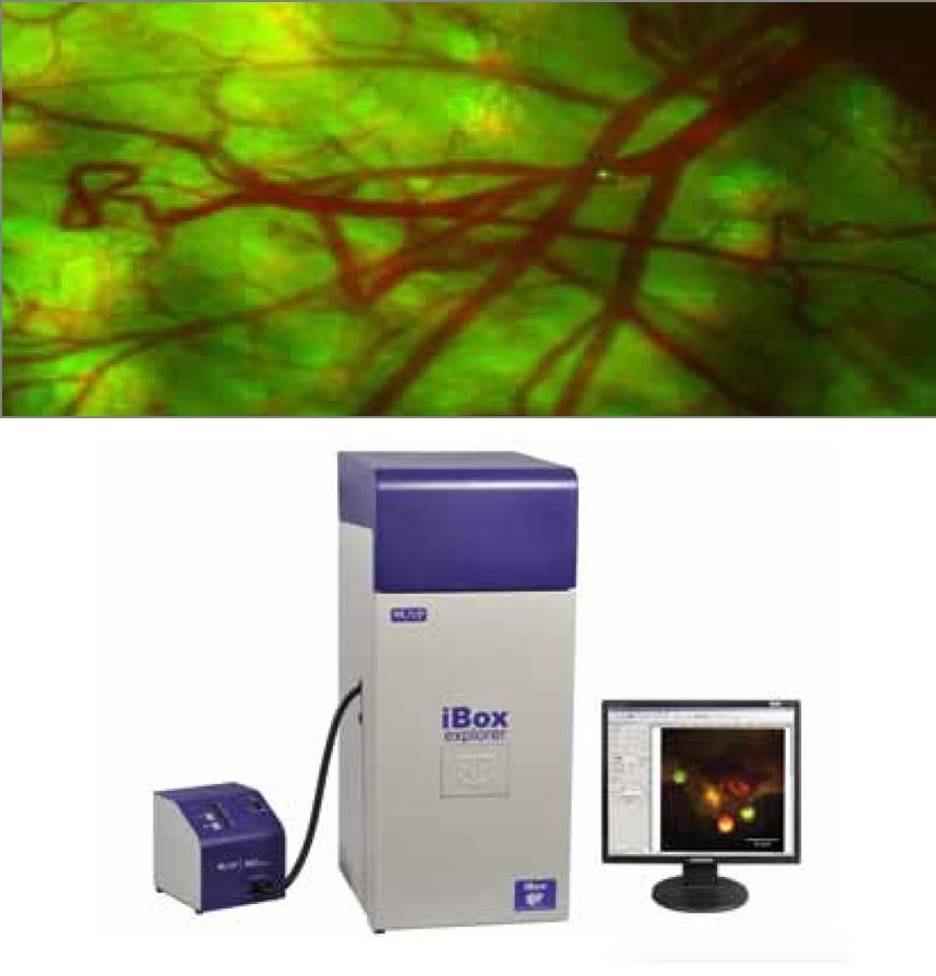
UVP is a leading manufacturer and developer of fluorescence and luminescence-based bio imaging systems for Genomic, Proteomic and In Vivo applications for pre-clinical markets. The latest addition to UVP’s iBox series of in vivo small animal imaging systems, iBox® Explorer™ Imaging Microscope, combines AntiCancer’s proprietary fluorescent-protein-based small-animal imaging technology with a UVP innovative world class imaging system. UVP and Edmund Optics® collaborated on developing system level specifications to meet the desired system improvements to enable researchers to visualize and capture multispectral fluorescently labeled cells. The required selected optical components were supplied by Edmund Optics®.
In Vivo fluorescence imaging of small animals has important implications for drug discovery, development in validating drug targets, and in studying the bio distribution, target binding, clinical effects, and toxicity of drug candidates. In Vivo studies include cancer research, heart disease, gene therapy and tumor detection using fluorescent tagged cells. Real time, non-invasive imaging capabilities speed up basic and preclinical research studies in oncology, cardiovascular and metabolic diseases. The main customers include pre-clinical biomedical research molecular diagnostics imaging labs, cancer research centers, diagnostic pathology labs, and oncology drug development research centers.
In Vivo imaging enables researchers to view biological processes at the molecular level. UVP’s new iBox® Explorer™ Fluorescence Imaging Microscope for In Vivo analysis is capable of visualizing whole organs as well as individual cells, making it well suited for the study of metastatic cancer and its high resolution and low light sensitivity allows detection of fluorescent markers deep within the animal. The demanding application performance requirements are achieved by engaging an intense broadband illumination using a powerful light source (UV to NIR), high sensitivity CCD camera with fast exposure times and an optimized filter set. The use of large-diameter optics with a large aperture for optimal light gathering assures long working distance and wide field-of-view from whole animal to cell-based resolution.


or view regional numbers
QUOTE TOOL
enter stock numbers to begin
Copyright 2023, Edmund Optics India Private Limited, #267, Greystone Building, Second Floor, 6th Cross Rd, Binnamangala, Stage 1, Indiranagar, Bengaluru, Karnataka, India 560038
California Consumer Privacy Act (CCPA): Do Not Sell or Share My Personal Information
California Transparency in Supply Chains Act